We provide expert gastroenterology care for patients with complex esophageal disorders
Baylor Scott & White Center for Esophageal Diseases is a specialized outpatient gastroenterology practice in Dallas focusing on esophageal disorders at Baylor University Medical Center, part of Baylor Scott & White Health.
Our center provides advanced care with a multidisciplinary team approach all while also conducting innovative research and participating in education at local, regional and national levels. Our providers are experts in the following esophageal disorders treatment:
- Achalasia and other motility disorders
- Barrett's esophagus
- Difficulty with swallowing
- Eosinophilic esophagitis
- Erosive esophagitis
- Esophageal strictures and narrowing
- Gastroesophageal reflux disease (GERD)
- Superficial esophageal cancer

Baylor Scott & White Center for Esophageal Diseases
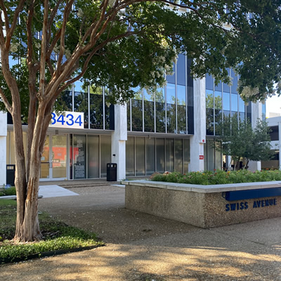
3434 Swiss Ave Ste 200, Dallas, TX, 75204
- Monday: 9:00 am - 5:00 pm
- Tuesday: 9:00 am - 5:00 pm
- Wednesday: 9:00 am - 5:00 pm
- Thursday: 9:00 am - 5:00 pm
- Friday: 9:00 am - 1:00 pm

Baylor Scott & White Center for Esophageal Diseases - Sammons Center (Prearranged Appointment)
3410 Worth St Ste 235, Dallas, TX, 75246
Insurances accepted
Baylor Scott & White has established agreements with several types of insurance to ensure your health needs are covered.
-
Aetna - (25)Choice POS IIOpen Access Managed ChoiceOpen Access Elect ChoiceAetna Medicare Eagle II (PPO)Aetna Medicare Choice Plan (PPO)STARSelectQPOSOpen Choice PPOOpen Access SelectManaged ChoiceAetna Medicare Dual Complete Plan (HMO D-SNP)Aetna Medicare Choice II Plan (PPO)Group Retiree Medicare PPO - Limited to Exxon/MobilAetna Signature AdministratorsHMOHealth Network OptionCHIPAetna Medicare Freedom Plan (PPO)Texas Preferred Plus IIHealth Network OnlyAetna Medicare Prime Plan (HMO)Aetna Medicare Eagle Plan (PPO)Aetna Medicare Freedom Preferred Plan (PPO)Aetna Medicare Value Plan (HMO)
-
American Health Advantage of Texas - (1)American Health Advantage of Texas HMO I-SNP
-
Baylor Scott & White Health Plan - (11)BSW Access PPOBSW Plus HMO-GroupBSW Plus HMO-Individual/FamilyBSW Plus PPO-GroupBSW Plus PPO-Individual/FamilyBSW Premier HMO-GroupBSW Premier HMO-Individual/FamilyBSW SeniorCare Advantage HMOBSW SeniorCare Advantage PPOBSWH Employee Network (SEQA & EQA)BSWH Employee Network Premium (PPO)/ HDHP
-
Blue Cross Blue Shield - (51)Blue Advantage - GoldTRS-ActiveCare Primary HDTRS-ActiveCare PrimaryTRS-ActiveCare 2Blue Advantage - SilverBlue Advantage Plus - BronzeBlue Cross Medicare Advantage Dual Care Plus (HMO SNP)Blue PremierFederal Basic OptionConsumer Directed HealthSelectBlue Essentials AccessBlue EssentialsTRS-Care StandardParPlanHigh Performance NetworkBlue Cross Group Medicare Advantage (PPO)Blue Cross Medicare Advantage (HMO)Blue Choice - Consumer Directed HealthSelect - Out-of-StateBlue Choice - TRS-Care StandardBlue Cross Medicare Advantage Dental Premier (PPO)Blue Choice - Texas A&MBlue Cross Medicare Advantage Choice Premier (PPO)Blue Cross Medicare Advantage Saver (HMO)Blue Essentials - HealthSelect of TexasBlue Cross Medicare Advantage Flex Access (PPO)Blue Choice - HealthSelect of Texas - Out-of-StateBlue Choice - TRS-ActiveCare 2Blue Cross Medicare Advantage Value (HMO)Blue Choice - City of DallasBlue Cross Medicare Advantage Choice Plus (PPO)Blue Advantage Plus - SilverFederal Standard OptionBlue Premier AccessBlue Cross Medicare Advantage Saver Plus (PPO)Blue Cross Medicare Advantage Dental Value (HMO)Blue ChoiceBlue Cross Group Medicare Advantage Open Access (PPO)STAR KidsCHIPBlue Essentials - Consumer Directed HealthSelectBlue Choice - TRS-ActiveCare HDBlue Advantage - BronzeBlue Cross Medicare Advantage Protect (PPO)Blue Advantage Plus - GoldBlue Essentials - TRS-ActiveCare PrimaryBlue Cross Medicare Advantage Health Choice (PPO)Blue Essentials - TRS-ActiveCare Primary+STARFederal FEP Blue FocusBlue Cross Medicare Advantage Flex (PPO)Blue Cross Medicare Advantage Classic (PPO)
-
Centivo Network - (1)Centivo Network - Baylor Scott & White Premier
-
Cigna - (17)Cigna Medicare AdvantageOpen Access Plus In-NetworkCigna Preferred Savings Medicare (HMO)NFL Dedicated Hospital Network ProgramCigna+OscarBSW Extended PPOChoice FundLocalPlusCigna True Choice Medicare (PPO)Open AccessCigna Preferred Medicare (HMO)Point of Service Open AccessLocalPlus In-NetworkCigna True Choice Courage Medicare (PPO)Cigna Preferred Savings Medicare (PPO)Cigna TotalCare (HMO D-SNP)Open Access Plus
-
Curative - (1)Curative
-
DFW ConnectedCare - (1)American Airlines Employee Benefit Plan
-
EHN - (1)Employers Health Network
-
FirstCare Health Plans - (1)CHIP
-
HealthSmart - (2)Accel NetworkPreferred Network
-
Humana - (9)ChoiceCareHumana PreferredHumanaChoice (PPO)Humana Honor (PPO)Humana Gold Choice (PFFS)National POSHumana USAA Honor with Rx (PPO)PPOHumanaChoice (Regional PPO)
-
Imagine Health - (1)Imagine Health Network
-
Nebraska Furniture Mart - (2)OnyxEmerald
-
Parkland Community Health Plan - (2)CHIPSTAR - HealthFirst
-
PHCS Network - (1)PHCS Primary PPO
-
ProCare - (1)Advantage Institutional Special Needs Plan (HMO POS I-SNP)
-
QuickTrip - (1)Employee Benefit Plan
-
Superior Health Plan - (18)Ambetter Core EPO - GoldWellcare Assist (HMO)Ambetter Core EPO - SilverWellcare Giveback (HMO)Wellcare by Allwell Complement Assist (HMO)Wellcare TexanPlus No Premium (HMO)Ambetter Core EPO - BronzeWellcare No Premium Rx Plus Open (PPO)Superior STAR+PLUS (MMP)Wellcare No Premium Open (PPO)Wellcare by Allwell Dual Access Harmony (HMO D-SNP)Wellcare by Allwell Dual Liberty Nurture (HMO D-SNP)STAR+PLUSWellcare Specialty No Premium (HMO C-SNP)Wellcare by Allwell Patriot No Premium (HMO)Wellcare Dual Access Open (PPO D-SNP)Wellcare Dual Access (HMO D-SNP)Wellcare Dual Liberty (HMO D-SNP)
-
TriWest HealthCare - (1)Community Care Network
-
United HealthCare - (32)AARP Medicare Advantage Walgreens (PPO)Navigate PlusEDGECoreAll SaversChoiceNavigateCore EssentialChoice PlusCharter PlusErickson Advantage Guardian (HMO-POS I-SNP)Nexus ACO - Referral RequiredErickson Advantage Freedom (HMO-POS)UnitedHealthcare Group Medicare Advantage (PPO)Erickson Advantage Signature with Drugs (HMO-POSSelectSelect PlusUnitedHealthcare Nursing Home Plan (PPO I-SNP)UnitedHealthcare Assisted Living Plan (PPO I-SNP)Charter BalancedSurestNavigate BalancedErickson Advantage Liberty with Drugs (HMO-POS)CharterNexus ACO - Open AccessErickson Advantage Liberty without Drugs (HMO-POS)Erickson Advantage Champion (HMO-POS C-SNP)OptionsSTAR KidsCHIPSTAR+PLUSSTAR
-
WellMed - (15)AARP Medicare Advantage SecureHorizons Plan 2 (HMO-POS)AARP Medicare Advantage (HMO-POS)AARP Medicare Advantage Choice (PPO)AARP Medicare Advantage SecureHorizons Plan 1 (HMO-POS)UnitedHealthcare Dual Complete Choice (Regional PPO DSNP)AARP Medicare Advantage Patriot (HMO-POS)UnitedHealthcare Dual Complete Select (HMO POS DSNP)UnitedHealthcare Dual Complete (HMO DSNP)AARP Medicare Advantage Walgreens (PPO)UnitedHealthcare Medicare Silver (Regional PPO CSNP)AARP Medicare Advantage (HMO)UnitedHealthcare Medicare Advantage Ally (HMO POS CSNP)UnitedHealthcare Group Medicare Advantage (HMO)UnitedHealthcare Medicare Gold (Regional PPO CSNP)UnitedHealthcare Medicare Advantage Choice (Regional PPO)
-
Tricare - (3)PrimeSelectExtra
We couldn’t find any results for ""
Diagnostic services
Colonoscopy
A colonoscopy is an endoscopic procedure to examine the inner lining of your large intestine, rectum and colon.
A thin, flexible tube called a colonoscope is inserted into the rectum and steered through the colon by the physician to look at the lining for any abnormalities.
Biopsies may be performed to sample the tissue.
If identified, polyps may be removed through the scope.
Endoflip
Endoflip is an assessment of the esophageal motility performed during a sedated endoscopic procedure.
The technology utilizes impedance planimetry with a functional luminal imaging probe to provide information on the dimensions and the pressure in the GI tract.
We use it to characterize function of the esophageal body and lower esophageal sphincter, help tailor treatments and gauge adequacy of interventions.
Endomicroscopy
Endomicroscopy provides real-time imaging of the gastrointestinal lining during an endoscopic procedure at the microscopic level.
Endomicroscopy may be used to better detect subtle abnormalities such as a precancerous change in the lining or outline the borders of a subtle lesion to plan therapy in real time.
Endomicroscopy systems that may be available include volumetric laser endomicroscopy and confocal laser endomicroscopy, and each offer different resolutions and field of view.
Endoscopic ultrasound
An endoscopic ultrasound is an endoscopic procedure that uses a specialized endoscope that has both a camera and an ultrasound at the end of the long flexible scope.
The ultrasound probe can image through the wall of the GI tract and adjacent organs in the chest and the abdomen.
A needle can be inserted to sample cells for analysis if needed.
Endoscopy
An upper endoscopy is a procedure that allows your gastroenterologist to examine the lining of your upper gastrointestinal tract.
A flexible, lighted tube is inserted through your mouth and into the esophagus, stomach and the first part of the duodenum.
Typically, patients are sedated for the procedure. During this procedure, the physician may take biopsies (small tissue samples for analysis).
In some cases, your physician may discuss additional diagnostic or therapeutic maneuvers may be performed during an upper endoscopy.
Esophagram
This is a radiology test to assess the structure and the overall motor coordination of the esophagus.
During this test, you swallow liquid barium, which outlines the structure of the esophagus.
The radiologist may also be able to comment if there is a major motor coordination problem that may prompt additional testing.
Some studies also include swallowing a barium pill as part of the examination.
You are awake for the procedure.
High-resolution manometry
High-resolution manometry is a diagnostic test performed to check the function of the esophagus and evaluate for a motility disorder of the esophagus.
High-resolution manometry measures pressures throughout your esophagus using a series of closely spaced pressure sensors on a thin catheter.
The thin catheter is inserted through the nose and runs down the esophagus into the stomach and is positioned to allow for the measurements throughout the esophagus.
You are awake for the procedure.
pH and impedance testing
A pH and impedance study is an outpatient test that measures the amount of acid or non-acid reflux of the stomach contents into the esophagus.
This is a catheter-based test. The catheter goes into your nose and is positioned along your esophagus with the end in the stomach.
The catheter is secured on the outside, and you are given a portable recording device.
The sensors along the catheter demonstrate the acid exposure, which reflects acid reflux and the fluid that comes up and may reflect weakly acidic or non-acidic reflux.
Wireless pH testing
Wireless pH testing allows your physician to evaluate the acid exposure in your esophagus while you continue your normal activities.
During an upper endoscopic procedure, the physician places a small capsule in your lower esophagus. The capsule records activity in that area for over a 48-hour period or 96-hour period and transmits acid levels to a wireless recording device which is worn on a belt.
You document meals and any symptoms during the study period. You return the recording device after the study period, and then your physician is able to download the data from the recording device and analyze the data to provide information about the severity of acid reflux.
The capsule will fall off on its own and does not need to be retrieved.
Therapeutic services
- Anti-reflux surgery/fundoplication
- Complex stricture dilation
- Cryotherapy ablation
- Endoscopic mucosal resection (EMR)
- Endoscopic submucosal dissection (ESD)
- Esophagectomy
- Heller myotomy (laparoscopic)
- Hybrid argon plasma coagulation (hybrid APC)
- Magnetic lower esophageal sphincter augmentation (LINX)
- Peroral endoscopic myotomy (POEM)
- Pneumatic balloon dilation
- Radiofrequency ablation (RFA)
- Revisional surgery after fundoplication and hiatal hernia repairs
Anti-reflux surgery/fundoplication
A surgical fundoplication is a surgical procedure to provide additional support the to lower esophageal muscles to reduce fluid from coming up from the stomach into the esophagus.
It is considered in some patients with acid reflux disease.
The surgeon wraps the top portion of the stomach around the junction of the esophagus and the stomach to reduce the reflux of contents back into the esophagus.
Complex stricture dilation
Some esophageal strictures may require multiple tools, multiple sessions or different approaches to provide improved caliber of the lumen.
Cryotherapy ablation
Cryotherapy ablation is a procedure that is performed during endoscopy to destroy the lining of the superficial diseased tissue with the cycle of freezing and thawing the tissue.
The therapy exposes cells to extreme cold and then allows cells to thaw to cause the death of the cells in the targeted area.
The goal is to treat by destroying the diseased tissue, and the lining that regrows is the normal esophageal lining.
Endoscopic mucosal resection (EMR)
Endoscopic mucosal resection (EMR) is a procedure that is performed during an endoscopy to remove the superficial tissue.
The physician may remove tissue through the scope in pieces that are larger and deeper than standard biopsy samples.
EMR can be used in superficial precancerous or cancerous disease in the gastrointestinal tract to both allow for a more accurate diagnosis and also for treatment by removing the diseased tissue.
Endoscopic submucosal dissection (ESD)
Endoscopic submucosal dissection is an endoscopic procedure that is used for superficial cancers of the GI tract. The physician uses endoscopic knives and specialized techniques to remove the lesion through the scope. The goal in appropriately selected patients is to perform a curative resection through the scope without the need for surgery.
Esophagectomy
Esophagectomy is a complex surgery in which the esophagus is removed due to esophageal cancer or severe damage due to perforation or end-stage achalasia. Reconstruction of a new food pipe is often performed with another organ such as the stomach or part of the large intestine.
Heller myotomy (laparoscopic)
A laparoscopic Heller myotomy is a minimally invasive surgical procedure used to treat achalasia.
This surgical procedure uses several small incisions to utilize small cameras and instruments to cut the muscle of the lower esophagus in patients with achalasia.
Hybrid argon plasma coagulation (hybrid APC)
Hybrid APC allows for tissue destruction in the GI tract with thermal energy delivered after a submucosal cushion is created to decrease deeper injury.
Magnetic lower esophageal sphincter augmentation (LINX)
Magnetic lower esophageal sphincter augmentation is a novel minimally invasive surgical procedure in which the surgeon places a ring of magnetic beads around the lower portion of the esophagus to create more support to reduce the reflux of contents back into the esophagus.
Peroral endoscopic myotomy (POEM)
Peroral endoscopic myotomy (POEM) is a novel therapeutic endoscopic procedure used to treat achalasia.
The procedure is a minimally invasive alternative to a surgical procedure that uses the endoscope to enter the wall of the esophagus to access and cut the muscle of the lower esophagus in patients with achalasia.
Pneumatic balloon dilation
Larger caliber balloon dilation is considered in the setting of treatment of achalasia and some post-surgical states. In addition to the standard pneumatic balloon dilations, we also have available the EsoFLIP dilation system which allows for incremental adjustments and real-time feedback of the dilation.
Radiofrequency ablation (RFA)
Radiofrequency ablation (RFA) is a procedure that is performed during endoscopy to destroy the lining of superficial diseased tissue with thermal energy.
Your physician may use a balloon catheter or different-sized probes depending on the area that needs to be treated.
The goal is to treat by destroying the diseased tissue and the lining that regrows is the normal esophageal lining.
Revisional surgery after fundoplication and hiatal hernia repairs
Sometimes an anti-reflux surgery may have unexpected symptoms or outcomes after surgery. Revisional surgery requires a thorough evaluation and are more complex than the initial surgery.
Pay bill
Baylor Scott & White Health is pleased to offer you multiple options to pay your bill. View our guide to understand your Baylor Scott & White billing statement.
We offer two online payment options:
- Make a one-time payment without registering by selecting the "Pay a Bill as a Guest" option.
- Enroll or login to your MyBSWHealth account to view account balances and statements, setup a payment plan or enroll in paperless statements.
Other payment options:
-
Pay by mail
To ensure that your payment is correctly applied to your account, detach the slip from your Baylor Scott & White billing statement and return the slip with your payment. If paying by check or money order, include your account number on the check or money order.
Please mail the payment to the address listed on your statement.
-
Pay by phone
Payments to HTPN can be made over the phone with our automated phone payment system 24 hours a day, seven days a week. All payments made via the automated phone payment system will post the next business day. Please call 1.866.377.1650.
If you need to speak to someone about a bill from a Baylor Scott & White Hospital, our Customer Service department is available to take payments over the phone from Monday through Friday from 8:00 AM - 5:00 PM and can be reached at 1.800.994.0371.
-
Pay in person
Payments can be made in person at the facility where you received services.
Financial assistance
At Baylor Scott & White Health, we want to be a resource for you and your family. Our team of customer service representatives and financial counselors are here to help you find financial solutions that can help cover your cost of care. We encourage you to speak to a team member before, during or after care is received.
Patient forms
To ensure that your visit to our office is as convenient and efficient as possible, we are pleased to offer our registration forms online. The patient registration form may be completed electronically and printed for better legibility or completed manually.
Other helpful information
Research
The Center for Esophageal Research is devoted to conducting innovative, translational research in a multidisciplinary setting in order to advance understanding of esophageal diseases and to improve the treatment of patients with those diseases.
Focus areas of translational research within the Center For Esophageal Research in Dallas include:
- Advanced endoscopic imaging of esophageal diseases
- Barrett’s esophagus
- Eosinophilic esophagitis
- Esophageal adenocarcinoma
- Esophageal squamous cell carcinoma
- Gastroesophageal junction adenocarcinoma
- Reflux esophagitis/gastroesophageal reflux disease (GERD)/non-erosive reflux disease (NERD)
- Swallowing disorders
Dr. Souza discussing how aspirin may help prevent Barrett's esophagus
Coordinators
Clinical studies coordinator
Daniel Kim
Histopathology coordinator
Elizabeth Cook, H.T. (ASCP)
Faculty
Faculty of The Center For Esophageal Research
- Stuart J. Spechler MD, Co-Director
- Rhonda F. Souza, MD, Co-Director
- Vani J.A. Konda, MD
- Qiuyang (Daniel) Zhang, Ph.D.
- Xi Zhang, MD
Physician publications
Yadlapati R, Vaezi MF, Vela MF, Spechler SJ, Shaheen NJ, Richter J, Lacy BE, Katzka D, Katz PO, Kahrilas PJ, Gyawali CP, Gerson L, Fass R, Castell DO, Craft J, Hillman L, Pandolfino JE. Management options for patients with GERD and persistent symptoms on proton pump inhibitors: recommendations from an expert panel. Am J Gastroenterology. 2018 Apr 24. [Epub ahead of print]
Mosher CA, Brown GR, Weideman RA, Crook TW, Cipher DJ, Spechler SJ, Feagins LA. Incidence of colorectal cancer and extracolonic cancers in veteran patients with inflammatory bowel disease. Inflammatory Bowel Disease 2018; 24:617-623.
Spechler SJ. Speculation as to why the frequency of eosinophilic esophagitis is increasing. Curr Gastroenterology Rep 2018; 20:26.
Spechler SJ. Cardiac Metaplasia: Follow, Treat, or Ignore? Dig Dis Sci 2018 Apr 18. [Epub ahead of print]
Spechler SJ, Katzka DA, Fitzgerald RC. New screening techniques in Barrett's esophagus: Great ideas or great practice? Gastroenterology 2018; 154:1594-1601.
Konda VJA, Spechler SJ. Endoscopic eradication therapy and the test of time. Gastrointestinal Endoscopy 2018; 87:85-87.
Huo X., Zhang X.. Yu C., Cheng E., Zhang Q., Dunbar K.B., Pham T.H., Lynch J.P., Wang D.H., Bresalier R.S., Spechler S.J., Souza R.F. Aspirin Prevents NF-kB Activation and CDX2 Expression Stimulated by Acid and Bile Salts in Oesophageal Squamous Cells of Barrett's Oesophagus Patients. Gut, 67: 606-615, 2018.
Choi S., Cui C., Luo Y., Kim S-H., Ko J-K., Huo X., Ma J., Fu L-W., Souza R.F.,Korichneva I., Pan Z. Selective Zinc Inhibitory Effect on Cell Proliferation in Esophageal Squamous Cell Carcinoma through Orail1. FASEB Journal, 32: 404-416, 2018.
Wang J., Park J.Y., Huang R., Souza R.F., Spechler S.J., Cheng E. Obtaining Adequate Lamina Propria for Subepithelial Fibrosis Evaluation in Pediatric Eosinophilic Esophagitis. Gastrointestinal Endoscopy, 87: 1207-1214, 2018.
Han J., Jackson D., Holm J., Turner K., Ashcraft P., Wang X., Cook B., Arning E., Genta R.M., Venuprasad K., Souza R.F., Sweetman L., Theiss A.L. Elevated D-2-Hydroxyglutarate during Colitis Drives Progression to Colorectal Cancer. PNAS, 30: 1057-1062, 2018.
Odiase E., Schwartz A., Souza R.F., Martin J., Konda V., Spechler S.J. New Eosinophilic Esophagitis Concepts Call for Change in Proton Pump Inhibitor Management Before Diagnostic Endoscopy. Gastroenterology, 154: 1209-1214, 2018.
Dellon ES, Liacouras CA, Molina-Infante J, Furuta GT, Spergel JM, Zevit N, Spechler SJ, Attwood SE, Straumann A, Aceves SS, Alexander JA, Atkins D, Arva NC, Blanchard C, Bonis PA, Book WM, Capocelli KE, Chehade M, Cheng E, Collins MH, Davis CM, Dias JA, Di Lorenzo C, Dohil R, Dupont C, Falk GW, Ferreira CT, Fox A, Gonsalves NP, Gupta SK, Katzka DA, Kinoshita Y, Menard-Katcher C, Kodroff E, Metz DC, Miehlke S, Muir AB, Mukkada VA, Murch S, Nurko S, Ohtsuka Y, Orel R, Papadopoulou A, Peterson KA, Philpott H, Putnam PE, Richter JE, Rosen R, Rothenberg ME, Schoepfer A, Scott MM, Shah N, Sheikh J, Souza RF, Strobel MJ, Talley NJ, Vaezi MF, Vandenplas Y, Vieira MC, Walker MM, Wechsler JB, Wershil BK, Wen T, Yang GY, Hirano I, Bredenoord AJ. Updated international consensus diagnostic criteria for eosinophilic esophagitis: Proceedings of the AGREE conference. Accepted, Gastroenterology, 2018. Epub.
Spechler SJ, Konda V, Souza RF. Can Eosinophilic Esophagitis Cause Achalasia and Other Esophageal Motility Disorders? Accepted, American Journal of Gastroenterology, 2018.
Agoston AT, Pham TH, Odze RD, Wang DH, Das KM, Spechler SJ, Souza RF. Columnar-Lined Esophagus Develops via Wound Repair in a Surgical Model of Reflux Esophagitis. Accepted, Cellular and Molecular Gastroenterology and Hepatology, 2018.
Souza R.F. and Spechler S.J. A New Candidate for the Progenitor Cell of Barrett's Metaplasia. Nature Reviews Gastroenterology and Hepatology, 15: 7-8, 2018.
Souza R. F., Rubenstein J, Kao J, Hirano I. Contributions from Gastroenterology: Acid Peptic Disorders, Barrett's Esophagus and Eosinophilic Esophagitis. Gastroenterology, 154: 1209-1214, 2018.
Souza R.F. Esophageal Adenocarcinomas: A Need for Speed Driven by Immune Pathways That Have Druggable Targets. Cellular and Molecular Gastroenterology and Hepatology, 5:652-653, 2018.
Konda V.J.A., Souza R.F. Biomarkers of Barrett's Esophagus: From the Laboratory to Clinical Practice. Digestive Diseases and Sciences, 63: 2070-2080, 2018.
Congratulations Dr. Spechler on being named the 2019 recipient of the AGA Institute Council Esophageal, Gastric and Duodenal Disorders (EGD) Section Research Mentor Award!
Research press
- Read Dr. Souza's published article on Barrett's esophagus
- Watch Dr. Spechler and Dr. Souza discuss their latest research on GERD
Congratulations Dr. Spechler on being named the 2019 recipient of the AGA Institute Council Esophageal, Gastric and Duodenal Disorders (EGD) Section Research Mentor Award! This award recognizes and expresses the appreciation of our section and AGA for your outstanding contributions to the mentoring and training of new investigators in the field.

